LinkExchange Member
The basic anatomical unit in the nervous system is a specialized cell called the neuron. One of the earliest descriptions of biological neurons is given in
There are at least 500 different types of biological neurons, but many neurons have a general structure similar to that described in this chapter. The following description of the function of biological neuron is simplified. However, at this level the elements of ANNs are usually modeled. A typical neuron is shown in the figure below.

Fig.1. A biological neuron
The principal components of a typical biological neuron are the cell body (soma) and the cell extensions. Many extensions of the single cell are long and filamentary; these structures are called processes. There are two different types of processes: the dendrites and the axon. A tree of dendrites is covered with special structures called synapses, where junctions are formed with other neurons. These synaptic contacts are the primary information processing elements in neural systems. Certain neurons are equipped with a long, specialized process called an axon. The axon is used for "digitizing" data for local transmission, and for transmitting data over long distances [I4, I20].
The function of the dendrites is the collection and conduction of electric potentials which are generated at the synapses when a presynaptic neuron experiences an action potential ("spike"). Temporal integration of signals occurs over the short term through charge storage on the capacitance of the cell membrane, and over the long term by means of internal second messengers and complex biochemical mechanisms. If the integrated intracellular potential in soma exceeds a certain value, called the threshold, an output is generated in the form of a nerve pulse - the action potential. This output pulse propagates down the axon, which ends in a tree of synaptic contacts to the dendrites of other neurons.
The resistance of a nerve's cytoplasm is sufficiently high that signals can not be transmitted more than about 1 millimeter before they are hopelessly spread out in time, and their information largely lost. For this reason, axons are equipped with an active amplification mechanism that restores the nerve pulse as it propagates. In lower animals, such as the squid, this restoration is done continuously along the length of the axon. In higher animals any axons are wrapped with a special insulating material called myelin, which reduces the capacitance between the cytoplasm and the extracellular fluid, and thereby increases the velocity at which signals propagate. The sheaths of these myelinated axons have gaps called nodes of Ranvier every few millimeters. These nodes act as repeater sites, where the signal is periodically restored. A single myelinated fiber can carry signals over a distance of 1 meter of more.
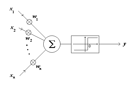
As mentioned above, signals are transmitted between neurons by electrical pulses travelling along the axon. These pulses impigne on the afferent neuron at synaopses. Each pulse occurring at a synapse initiates the release of a small amount of chemical substance or neurotransmitter which travels across the synaptic cleft and which is then received at post-synaptic receptor sites on the dendritic side of the synapse. The neurotransmitter becomes bound to molecular sites here which, in turn, initiates a change in the dendritic membrane potential. This post-synaptic-potential (PSP) change may serve to increase (hyperpolarise) or decrease (depolarise) the polarisation of the post-synaptic membrane. In the former case, the PSP tends to inhibit generation of pulses in the afferent neuron, while in the latter, it tends to excite the generation of pulses. The size and type of PSP produced will depend on factors such as the geometry of the synapse and the type of neurotransmitter. Each PSPwill travel along its dendrite and spread over the soma, eventually reaching the base of the axon (axon-hillock). The afferent neuron sums or integrates the effects of thousands of such PSPs over its dendritic tree and over time. If the integrated potential at the axon-hillock exceeds a threshold, the cell "fires" and generates an action potential or spike which starts to travel along its axon. This then initiates the whole sequence of events again in neurons contained in the efferent pathway. This functionality is captured in the artificial neuron known as the Threshold Logic Unit (TLU) originally proposed by McCulloch and Pitts in 1943. [I4, I5]. Below is a view of the TLU.

Fig.2. A Threshold Logic Unit
The TLU operation can be described as follows:
Let v denotes the internal activity level of the TLU and y is its output level. Then:
|
|
(1) |
|---|---|
|
|
(2) |
where:
| x | is the ith input signal to the TLU, |
|---|---|
| w | is the weight of the ith process of the TLU, |
| y | is the output of the TLU, |
| n | is the number of inputs of the TLU. |
Then, the TLU's output:
| (3) | ||
| (4) |
The value ![]() is refered to as a threshold. Considering that the TLU has only binary inputs, this extremely simple artificial neuron can calculate Boolean functions. However, this simple model of the biological neuron is not very interesting. What makes the neuron model more useful is its interconnection with other TLUs.
is refered to as a threshold. Considering that the TLU has only binary inputs, this extremely simple artificial neuron can calculate Boolean functions. However, this simple model of the biological neuron is not very interesting. What makes the neuron model more useful is its interconnection with other TLUs.
Please send Questions, Comments, Suggestions to
bencr@hotmail.comCopyright © 1997, 98 Rado Benc